02/08/2024
Have you ever wondered why ECG tests are vital for your heart health? An electrocardiogram (ECG) is a simple, non-invasive test that records your heart’s electrical activity. It can provide crucial insights into your heart health and detect conditions early on, which is key for effective treatment and management.
By understanding how your heart works and what an ECG can reveal, you can take proactive steps to maintain your heart health. Early detection of heart conditions such as arrhythmias can significantly improve outcomes and potentially save lives. Whether you are experiencing symptoms like palpitations, dizziness, or taking preventive measures, an ECG test can offer valuable information about your heart’s rhythm and function.
In this article, we’ll explore the basics of heart physiology, how the heart’s electrical system works, and the different types of arrhythmias that an ECG can detect. Let’s dive in and see what your heart might be trying to tell you.
Physiology of the Heart
To understand how an ECG works, let’s break down some basics of the heart’s physiology. The heart consists of four chambers: the two upper chambers called atria, and the two lower chambers called ventricles. The atria receive blood from the body and lungs, while the ventricles pump it out to the lungs and the rest of the body3.
Blood enters the right atrium from the body – if you think of your own heart, it’s the one on your right – moves to the right ventricle and is pumped to the lungs for oxygenation. Oxygen-rich blood then flows into the left atrium, down to the left ventricle, and is pumped out to supply the body. This continuous cycle ensures that your body receives the oxygen and nutrients it needs to function properly3.
Electrical System of the Heart
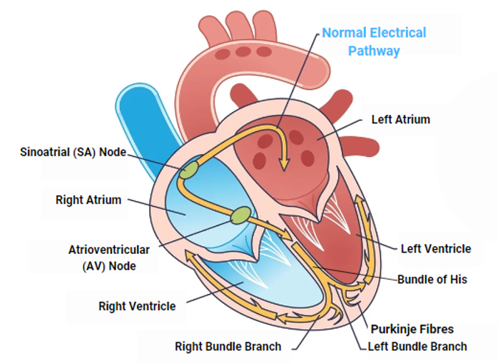
Imagine the heart as an electrical circuit. The heart’s electrical impulses start in the sinoatrial (SA) node, often called the natural pacemaker, located in the right atrium. These impulses then travel through the atria to the atrioventricular (AV) node, which acts like a gateway, slowing down the impulses before they move to the ventricles. From the AV node, the impulses travel through the Bundle of His, a collection of specialised heart muscle cells responsible for transmitting electrical impulses from the atria to the ventricles. The Bundle of His is an integral part of the heart’s electrical conduction system and ensures that the impulses are conducted rapidly and efficiently. It then divides into right and left bundle branches, which carry the impulses down through the septum separating the ventricles. Finally, the impulses reach the Purkinje fibres, causing the ventricles to contract and pump blood out of the heart4,5.

Understanding Arrhythmias
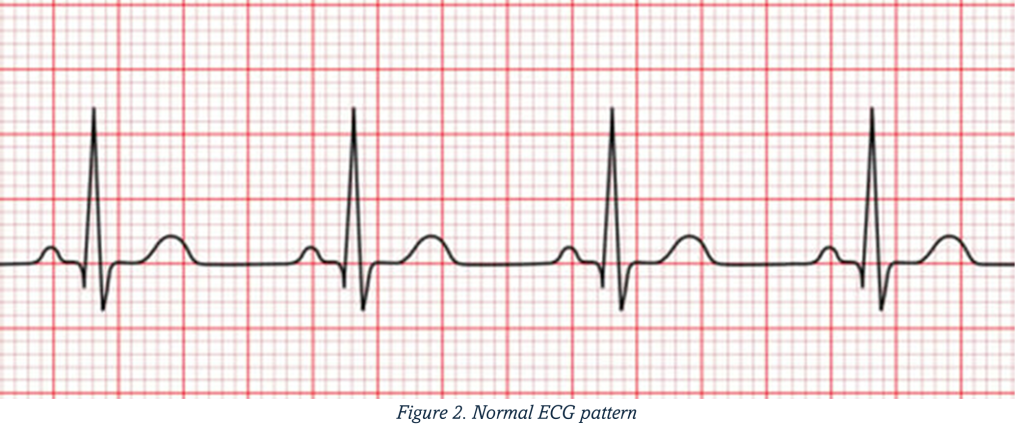
An ECG helps you understand your heart’s health by measuring its electrical activity. By measuring the electrical activity of your heart, an ECG can provide a snapshot of your heart’s rhythm and help detect any irregularities or arrhythmias. Figure 2 shows a normal ECG, which represents the way electrical signals move through the heart. This ensures the heart’s chambers contract in a coordinated manner, allowing efficient blood flow from the atria to the ventricles and then to the rest of the body.

Atrial Fibrillation
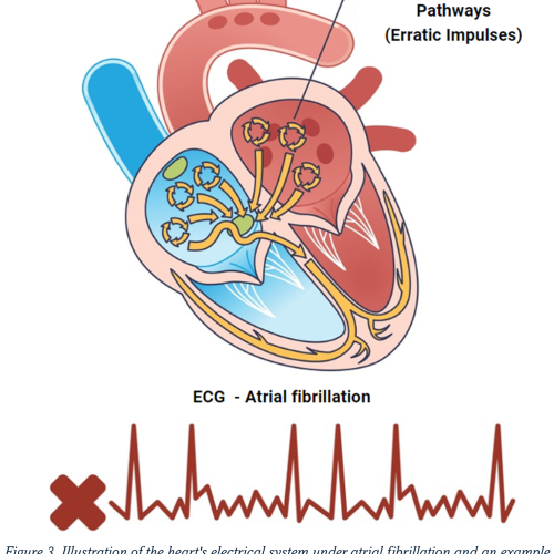
Atrial fibrillation (AF) is the most common type of arrhythmia6 occurs when the upper chambers of your heart beat irregularly and rapidly, making your heart feel like it’s fluttering. This irregularity causes the atria to quiver or fibrillate instead of contracting effectively, leading to poor blood flow from the atria to the ventricles. AF can cause symptoms such as heart palpitations, shortness of breath, fatigue, and an increased risk of stroke and heart failure due to the potential for blood clots forming in the atria and traveling to the brain or other parts of the body7.

Tachycardia
Tachycardia is when your heart beats faster than the normal range at rest, typically over 100 beats per minute. Causes can include stress, exercise, or medical conditions like AF. Symptoms might include palpitations, dizziness, or chest pain. If left untreated, tachycardia can lead to more serious heart issues6.
Tachycardia is normally the result of an additional electrical pathway either within the AV node (AV nodal re-entrant tachycardia, AVNRT) or outside the AV node (Wolff-Parkinson-White syndrome, WPW). In AVNRT, there is a small extra pathway near the AV node, creating a re-entrant circuit. This causes the heart to beat more than 100 times a minute8.
Similarly, in WPW syndrome, an accessory pathway known as the Kent bundle connects the atria and ventricles, allowing electrical signals to bypass the AV node9. This results in a continuous feedback loop where the electrical signal travels the normal pathway from the atria to the ventricles but returns via the additional pathway, initiating a premature subsequent heartbeat, leading to an abnormally fast heartrate.
In other cases, rather than an extra pathway, there is an abnormal ‘focus’ – a specific area in the heart that independently generates electrical impulses. This focus acts as a second natural pacemaker, causing the heart to beat faster than the SA node would normally allow4.
Bradycardia
Bradycardia is when your heart beats slower than normal, typically less than 60 beats per minute at rest. It can be caused by aging, heart disease, or medication. Symptoms might include fatigue, dizziness, or fainting. While bradycardia can be normal in some people, especially athletes, it can also be a sign of a problem with the heart’s electrical system6.
A common cause of bradycardia is heart block, which occurs when the electrical signal begins in the SA node but is delayed or blocked before it reaches the ventricles. This results in an inadequate heartbeat and pumping action, causing symptoms such as fatigue, dizziness, and fainting10,11.
Another cause of bradycardia is SA node dysfunction – when the SA node does not work with its usual regularity, causing a slow heart rate. This can result from age-related wear and tear, heart disease, or certain medications. SA node dysfunction often presents with similar symptoms and requires careful management12.
Importance of ECG Tests.
So, why are ECG tests important? They’re crucial for detecting various heart conditions early on, which is vital for effective treatment and management. ECG tests can reveal problems like arrhythmias, heart attacks, and other heart diseases. Early detection through regular ECG tests can lead to better management and treatment of these conditions, potentially saving lives1,2.
Preventative Heart Health
Regular ECG tests are essential for proactive heart health management. By detecting issues early, you can take steps to manage your heart health more effectively. Alongside regular ECG tests, maintaining a healthy lifestyle is key. This includes eating a balanced diet, exercising regularly, avoiding smoking, and managing stress1. At Randox Health, we offer a range of diagnostic tests and health services to help you keep your heart in top condition.
Randox Health ECG Test
At Randox Health, our comprehensive ECG test is designed to provide accurate and reliable results. Our test is non-invasive, quick, and performed by trained professionals. What sets our ECG test apart is our commitment to accuracy and thorough analysis, which we provide in our detailed and easy-to-follow reports.
Getting an ECG test at Randox Health is simple and convenient. Here’s how it works:
- Booking: You can easily book an appointment online or by phone.
- Preparation: On the day of the test, there’s no special preparation needed. Just wear comfortable clothing.
- The Test: During the test, small electrodes will be placed on your chest, arms, and legs to record the electrical activity of your heart.
- Results: The test itself takes about 10 minutes. Afterward, you’ll get a detailed report by midnight the following day, explaining your results and any further steps you might need to take.
Frequently Asked Questions (FAQs)
Is this test painful?
No, an ECG test is completely painless. You might feel a slight discomfort when the electrodes are removed, but that’s it.
Do I need to fast before the test?
No fasting is required for an ECG test
How often should I get an ECG test?
This depends on your individual health and risk factors. It’s best to consult with a healthcare professional to determine what’s right for you.
Conclusion
In summary, an ECG test is invaluable for monitoring and maintaining heart health. Take a proactive step towards better heart health with Randox Health today.
The Randox Health ECG test offers a reliable and accurate way to detect heart conditions early. By understanding your heart’s physiology and electrical system, you can better appreciate the importance of regular ECG tests. So, why wait? Take a proactive step towards better heart health with Randox Health today.
Glossary
Atria: The two upper chambers of the heart that receive blood from the body and lungs.
Atrial Fibrillation (AF): A common type of arrhythmia where the atria (upper chambers of the heart) beat irregularly and often rapidly.
Arrhythmia: Any condition in which the heart beats with an irregular or abnormal rhythm.
Atrioventricular (AV) Node: A part of the electrical conduction system of the heart that coordinates the top of the heart. It electrically connects the atria and ventricles.
AV Nodal Re-entrant Tachycardia (AVNRT): A type of tachycardia that involves an extra pathway near or within the AV node, creating a re-entrant circuit.
Bradycardia: A condition where the heart rate is slower than normal, typically less than 60 beats per minute.
Bundle of His: An integral part of the heart’s electrical conduction system and ensures that the impulses are conducted rapidly and efficiently.
ECG (Electrocardiogram): A non-invasive test that measures the electrical activity of the heart to help detect heart conditions.
Heart Block: A condition where the electrical signal begins in the SA node but is delayed or blocked before it reaches the ventricles.
Kent Bundle: An abnormal extra conduction pathway between the atria and the ventricles that can cause rapid heart rhythms.
Purkinje Fibres: Specialised conductive fibres located within the walls of the ventricles that help propagate the electrical impulse to the heart muscle, causing it to contract.
Sinoatrial (SA) Node: Often referred to as the heart’s natural pacemaker, it is located in the right atrium and initiates the electrical impulses that set the pace for the heart rate.
Tachycardia: A condition where the heart rate is faster than normal, typically over 100 beats per minute.
Ventricles: The two lower chambers of the heart that pump blood to the lungs and the rest of the body.
Wolff-Parkinson-White (WPW) Syndrome: A condition where there is an extra electrical pathway between the atria and ventricles, known as the Kent bundle, leading to episodes of tachycardia.
References
- American Heart Association. Electrocardiogram (ECG or EKG). https://www.heart.org/en/health-topics/heart-attack/diagnosing-a-heart-attack/electrocardiogram-ecg-or-ekg.
- NHS. Electrocardiogram (ECG). https://www.nhs.uk/conditions/electrocardiogram/.
- Cleveland Clinic. Blood Flow Through the Heart. https://my.clevelandclinic.org/health/articles/17060-how-does-the-blood-flow-through-your-heart.
- USCF Benioff Children’s Hospital. The Heart’s Electrical System. https://www.ucsfbenioffchildrens.org/education/the-hearts-electrical-system.
- John Hopkins Medicine. Anatomy and Function of the Heart’s Electrical System. https://www.hopkinsmedicine.org/health/conditions-and-diseases/anatomy-and-function-of-the-hearts-electrical-system.
- NHS. Arrhythmia. https://www.nhs.uk/conditions/arrhythmia/.
- Mayo Clinic. Atrial fibrillation. https://www.mayoclinic.org/diseases-conditions/atrial-fibrillation/symptoms-causes/syc-20350624.
- Mayo Clinic. Supraventricular tachycardia. https://www.mayoclinic.org/diseases-conditions/avnrt/cdc-20355254.
- NHS. Wolff-Parkinson-White syndrome. https://www.nhs.uk/conditions/wolff-parkinson-white-syndrome/.
- Mayo Clinic. Bradycardia. https://mayoclinic.org/diseases-conditions/bradycardia/symptoms-causes/syc-20355474.
- Cleveland Clinic. Sinus Bradycardia. https://my.clevelandclinic.org/health/diseases/22473-sinus-bradycardia.
- Mitchell LB. Sinus Node Dysfunction. https://www.msdmanuals.com/home/heart-and-blood-vessel-disorders/abnormal-heart-rhythms/sinus-node-dysfunction.


